glaucoma

5 Signs Your Pet Has an Eye Emergency

5 Signs Your Pet Has an Eye Emergency

It's not uncommon for a veterinary clinic to field plenty of questions concerning pet eye problems on the daily. “Watchful waiting” is advised with some minor complaints, but never with eyes.

A non-vet pet owner cannot assess an eye problem’s severity, and a description over the phone just doesn’t cut it. In other words, when someone calls in and says their pet’s eye “looks funny,” it’s time for a trip to the vet.

Your vet may even direct you to a board-certified veterinary ophthalmologist for immediate assessment if the emergency requires it.

Red, inflamed or swollen eyes? Each of these symptoms can be caused by different ailments.

1. Red Eye

The white of the eye should be pristine white, with the odd lazy blood vessel meandering across the surface. If you’re not sure what this looks like, check out your own eye in the mirror.

Red eyes are not normal.

Gently lift the upper eyelid to check, and you’ll see anything from a rosy pink to a livid red. As a rule of thumb, the angrier the eye looks, the more urgently it needs checking.

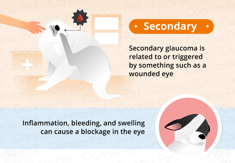
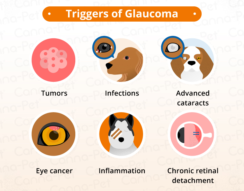
Causes of red-eye range from irritation and infection to a condition called glaucoma.

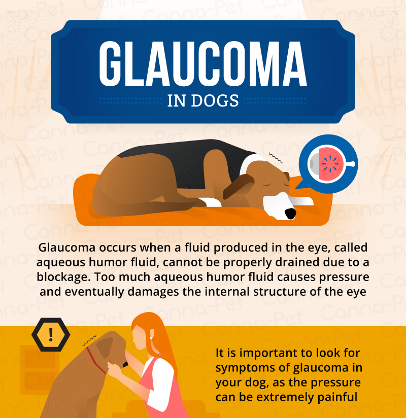
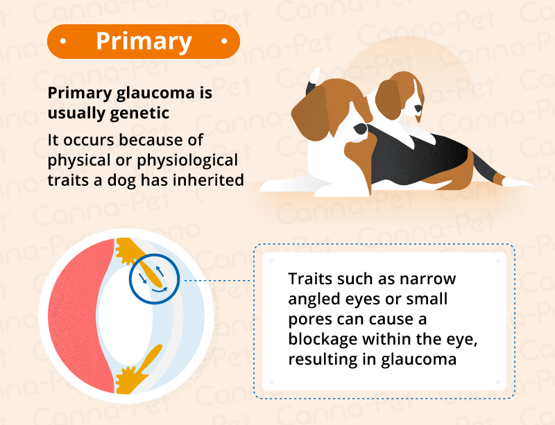
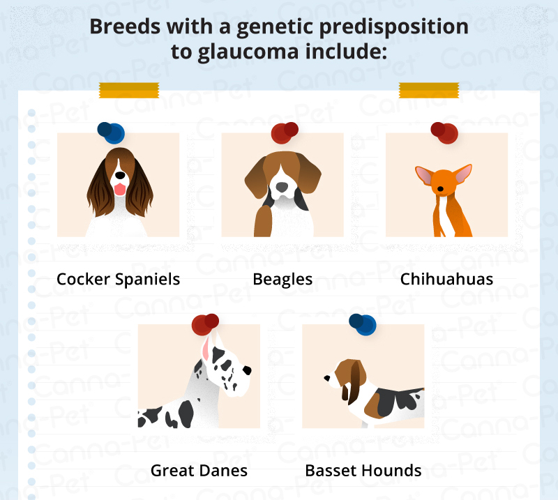
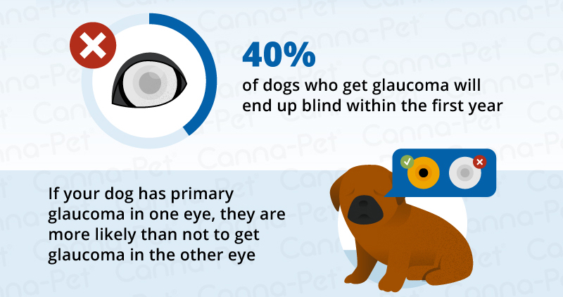
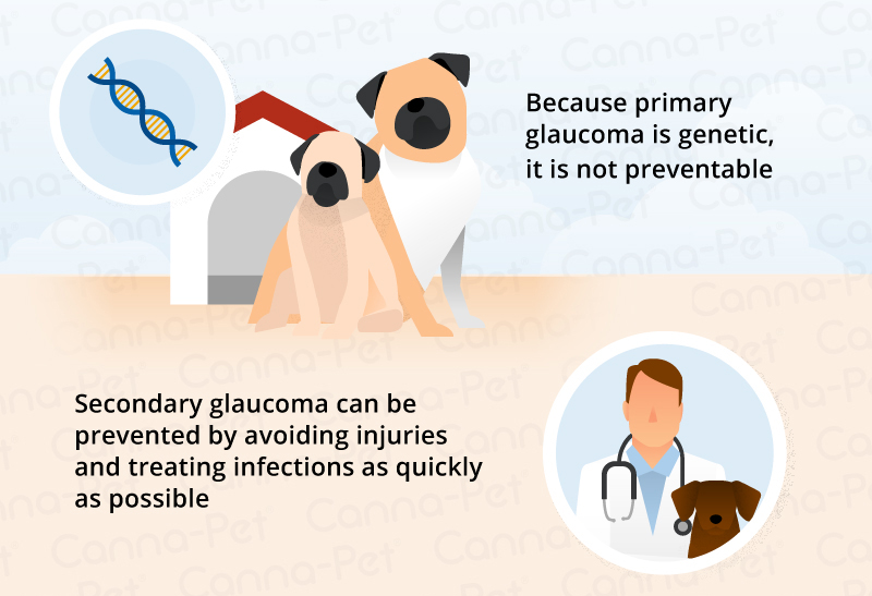
Glaucoma: Pressure builds within the eye, like blowing too much air into a balloon. The most common cause is a problem within the eye and is often breed-related (Basset Hounds, Cocker Spaniels, terriers and northern breeds are at greatest risk). The pressure can damage the retina and cause blindness, so swift action is essential.

Conjunctivitis: Infection causes reddening of the eye. Sometimes the problem can be self-limiting, but — especially if there is a sticky yellow-green discharge, too — please see a vet.

2. Yellow-Green Discharge

It’s normal to have a white gloop in the corner of an eye first thing in the morning. Just wipe this away with a clean, damp cotton towel.

Also, rust-colored gloop is fine. This is a normal gloop that’s been exposed to the air for a while and oxidized (like when you cut an apple in half and it goes brown).

What isn’t normal is a thick, yellow-green discharge from one or both eyes. This is commonly a sign of infection.

Have the vet check the eye because some infections need antibiotics, while others can occur as a complication of another problem that needs attention.

3. Swollen Eye

If there’s something odd about your pet’s face, compare one eyelid with the other to see if one side is swollen. Eyelid swelling can be the result of an allergy, trauma, or infection. It’s best to seek vet attention because the eyeball needs checking to make sure it wasn’t damaged.

4. Dull Eye

Is the eye lackluster? A normal eye is bright, and you can see reflections on the surface— but sometimes the surface is dull and reflections aren’t clear, or those images are broken up or haphazard.

The most common reasons for this are either a dry eye or a corneal ulcer:

Dry eye: Our eyes are kept comfortable by the production of tear fluid. A pet with dry-eye fails to produce enough tear fluid, which leads to the surface drying out. One consequence is a dull surface, and another is the eye tries to protect itself by producing a thick, glue-like discharge. In the long term, scar tissue forms, impairing the vision.

Corneal ulcer: This is like a burst blister on the surface of the eye. In some cases, it heals on its own, but other times it can be dangerous and cause perforation of the eye.

5. Closed Eye

A closed eye is painful: Just think of the last time you had grit in your eye.

The pain might be due to a corneal ulcer, a knock to the eye, or a foreign body — anything from dust to grass particles or even a twig. When our pets are sniffing around, it’s not uncommon to get something lodged behind the eyelids.

Your vet will put drops of local anesthetic into the eye to get a better look and remove the object.


5 Most Common Eye Emergencies (in Dogs)

Here, we go into more detail about some of the top ophthalmologic emergencies we see with dogs in general practice.

1. Corneal Ulcers

Corneal ulcers are the most common acute eye problems seen. (This goes for cats as well.) The cornea is the outer “skin” of the eye; an ulcer occurs if this layer has been damaged.

Corneal ulcers are often traumatic in origin, although certain diseases of the cornea can result in an ulcer. Trauma to the cornea can occur with a scratch from a bush, a stick or even another critter.

Treatment involves early diagnosis of the severity of the ulcer and the administration of appropriate eye medications.

More serious ulcers may require surgery, and frequent re-checks are needed to ensure the cornea is healing nicely. When a corneal ulcer goes from bad to worse, the cornea can actually rupture — this is a true emergency indeed.

2. Proptosis

Proptosis occurs when an eye literally bulges out of the socket and the eyelids entrap the globe. This occurs most frequently in brachycephalic breeds and is a true emergency.

Even with immediate care, the dog may lose the eye depending on the extent of trauma sustained by the extraocular muscles, nerves, and blood supply.

Many of these eyes can be replaced and vision saved in about 20% of dogs, but only if you act immediately.

3. Corneal Laceration

Corneal laceration occurs when there is a complete tear through the cornea. The most common offender from a cat claw.

A sharp object, like a stick, can also puncture the cornea. The dog is almost always holding the eye completely shut and is in significant pain. Again, immediate surgery may save the eye.

Get to the vet. There is a very small window of opportunity when it comes to repairing these eye injuries.

4. Lens Luxation

Lens luxation is over-represented in certain breeds like the Russell Terrier and other terriers. In these dogs, due to a genetic disorder, the lens can spontaneously luxate, or become dislocated. In other breeds, causes may vary — head trauma is one example. This is a difficult diagnosis and may require a visit to an ophthalmologist.

Compared with the normal eye, a lens luxation can look like a very dilated pupil or a blue or whitish eye. Removing the lens can save the eye and save the dog from pain and total blindness.

5. Acute Glaucoma

Acute glaucoma looks like a discolored, a “red” or an inflamed eye. There may be discharge and painful blinking (called blepharospasm).

Glaucoma occurs when, for whatever reason, the pressure in the eye elevates, leading to pain, secondary changes, and blindness. The condition is usually obvious in just one eye, but both eyes are at risk.

Again, this is over-represented in certain breeds like the Cocker Spaniel, Basset Hound, Shih Tzu, Great Dane, and northern breeds.
As with every other eye problem discussed here, acute glaucoma should be assessed and treated immediately. Both systemic (oral) and ophthalmic drugs are used. Get an opinion from a veterinary ophthalmologist to help you and your primary care vet manage the case.

Don’t mess around with the eyes. Ophthalmic problems need early diagnosis and treatment.


Hear From Us Again

Don't forget to subscribe to our email newsletter for more recipes, articles, and clinic updates delivered straight to your e-mail inbox.

Related Categories:

DOG HEALTH

CAT HEALTH

Glaucoma in Dogs: Causes, Signs & Treatment

blog_ Glaucoma _banner.png

Disease of the Optic Nerve in Dogs

Glaucoma is a condition in which pressure is placed on the eye, causing inadequate fluid drainage in the eye. If the condition becomes chronic or persists without treatment, it will eventually cause permanent damage to the optic nerve, resulting in blindness.

Glaucoma is common in certain dog breeds that are genetically predisposed, such as samoyeds, cocker Spaniels, poodles, chow chows, and Siberians. Unfortunately, 40% of dogs affected by glaucoma will become blind in the affected eye within the first year, regardless of medical or surgical treatment.


Symptoms and Types of Glaucoma

There are two main types of glaucoma: primary and secondary. Symptoms for sudden primary disease, due to the eye's inability to drain through the filtration angles of the eye, are as follows:

  • High pressure within the eye

  • Blinking of the eye

  • The eyeball may recede back into the head

  • Redness of the blood vessels in the whites of eyes

  • Cloudy appearance at front of the eye

  • Dilated pupil – or pupil does not respond to light

  • Vision loss


Long-term, advanced disease:

  • Enlargement of the eyeball (buphthalmos)

  • Obvious loss of vision

  • Advanced degeneration within the eye


Symptoms for secondary glaucoma, or glaucoma due to secondary eye infection(s), include:

  • High pressure within the eye

  • Redness of the blood vessels in the whites of eyes

  • Cloudy appearance at front of the eye

  • Inflammatory debris visible in the front of the eye

  • Possible constriction of the pupil

  • Possible sticking of the iris to either the cornea or the lens

  • Possible that the edge of the iris circularly sticks to the lens


In addition, there may be:

  • Headaches, with head pressing to relieve feelings of pressure in head

  • Loss of appetite

  • Change in attitude, less desire to play or interact


Causes

High pressure in the eye occurs when the normal outflow of fluid in the eye is impaired due to a primary eye disease such as the improper development of the eye's filtration angles, or secondary to other eye diseases such as primary lens luxation (slipping of the lens in the eye), inflammation of the tissues of the eye, eye tumor(s), or blood collection in the front of the eye from injury. In dogs, secondary glaucoma is more common than primary glaucoma.

Diagnosis 

You will need to give a thorough history of your dog's health, onset of symptoms, as far as you have been able to tell, and possible incidents that might have preceded this condition, such as injuries to the eye (even those which you consider minor). During the physical examination, your veterinarian will test the pressure within your dog's  eyes using a tonometer on the surface of the eye. If the disease began suddenly, your veterinarian will refer your dog to a veterinary ophthalmologist for a detailed examination of both eyes, including evaluation of the filtration angles by gonioscopy -- measuring the anterior of the eye. Pressure within the eye can measure as high as 45 to 65 mmHg, making this a very painful condition.

Electroretinography will be also performed by the veterinary ophthalmologist to determine if the eye will remain blind despite treatment. In secondary diseases, X-rays and an ultrasound may show abnormalities within the eye.

Often both eyes are affected, but not always. In cases where only one eye is affected, steps will be taken to protect the unaffected eye from developing a diseased condition.


Treatment

It is helpful to think of treating glaucoma like a major medical emergency. The faster and earlier you address the problem, the least likely your pet is to suffer maximum, sustained damages. The top three goals are to reduce pain, drain excess fluid, and reduce how much aqueous humor the eye produces.

Most treatments are geared toward pain management to help ward off the headaches associated with glaucoma, and in delaying or preventing the disease from happening in the second eye. Your vet may recommend performing a gonioscopy to determine your best shot at saving the remaining eye.

Reduce aqueous humor fluid.

Some treatment options seek to decrease the amount of fluid the eye produces, since draining the eye and keeping it drained can be tough, if not impossible in animals. There are pills and eye drops used for these purposes, and although they are helpful, they aren’t that great as a long-term option, and are ineffective in an emergency. The medications are most useful as a Band-Aid method until surgery of the affected eye is possible. This combination of treatment methods is usually the pet owner’s best chance at saving their dog’s vision.

Reduce stress.

As with humans, stress can be very detrimental to a dog’s health. The immune system cannot do its job and the body cannot fight off oxidative damage if stress levels are high.

Avoid treating a blind eye.

Medical treatment of an eye that’s already blind is not recommended. There’s no point in paying for expensive drugs and therapies to try to recover sight that is impossible to recover. In some cases, it might just be best to remove the blind eye entirely in order to mitigate pain and discomfort. It’s cheaper than the cost of medications and ongoing visits with the vet for eye exams.


Hear From Us Again

Don't forget to subscribe to our email newsletter for more recipes, articles, and clinic updates delivered to your inbox (here). Or, you can keep up to date by liking and following our Facebook page (here).

Related: We have more information under our dog health tags.

8 Ways to Help a Blind Cat

blog_ Blind Cat _banner.png

A blind cat can have a wonderful, happy life. It is not at all uncommon for pets, particularly older ones, to suffer vision loss. Normal cat vision is close to humans or perhaps just a little less. Pets have more problems focusing on near objects than people do, though, which is why your kitty may have some trouble seeing the last few kibbles in the food bowl.

Just as people over the age of 40 tend to need reading glasses, the same vision changes start to develop in almost all cats over the age of five. This normal change, called nuclear sclerosis, results in less flexibility of the lens, a hazy appearance, and less ability to focus on close objects. Pets still see pretty well despite the bluish tint to their eyes. Blind cats typically are still very happy. They continue to enjoy and remain engaged in life and the world around them—including their humans. Kitties do not need to see you to love you.


Your blind pet’s comfort level, safety, and emotional health are important. Follow these tips to keep the cat happy and comfortable.

Do not move food or litter box.

It is vital to keep the food, water bowls, litter box, and pet beds in the same spot, so your cat can easily find belongings.

Do some scent marking.

It may be helpful to “scent” important objects for the cat with strong odors such as peppermint to help its nose “see” what it is looking for.

Avoid rearranging the furniture.

Blind pets memorize and "mind-map" the house, and moving things around will confuse the cat. It is not at all unusual for a blind cat, for instance, to still insist on making floor-to-counter leaps with confidence as long as its memory remains fresh and accurate.

Safeguard dangerous zones.

Pad the sharp edges of furniture with bubble wrap until your cat learns to avoid the danger. Block off steep stairways with baby gates to prevent falls.

Use your voice to guide your cat.

Your pet’s personality and behavior may change a bit as vision fades. Some pets become more dependent on the owner, and act “clingy”—basically they will treat you as a guide, stand very close, and follow you around. Get in the habit of speaking to your cat when you enter or leave a room to help it keep track of your whereabouts.

Attach a bell to other animals in the house.

In multiple pet homes, another cat or dog may serve as a guide for the blind pet. Help your blind pet by attaching a bell or other noisemaker to the other animal's collar.

Create a safe spot in each room.

To avoid tripping over the pet that is always underfoot, provide a safe, comfy bed in each room. Very social cats may become standoffish once vision fades. They may want to avoid contact with houseguests to avoid being stepped on.

Don't startle your cat.

Blind pets also startle more easily, so always speak to your cat before petting him to avoid being accidentally nipped or swatted in reflex.


If you suspect your pet is sick, call your vet immediately. For health-related questions, always consult your veterinarian, as they have examined your pet, know the pet's health history, and can make the best recommendations for your pet.


Hear From Us Again

Don't forget to subscribe to our email newsletter for more recipes, articles, and clinic updates delivered to your inbox (here). Or, you can keep up to date by liking and following our Facebook page (here).

Related: We have more information under our cat health tags.